
The Placenta — Lifeline Between Mother and Fetus
After implantation, the trophoblast develops into the placenta — a unique organ that forms the interface between the mother and the developing fetus. The placenta is essentially temporary — it forms during pregnancy and is delivered after the baby (as the "afterbirth").
What is the placenta?
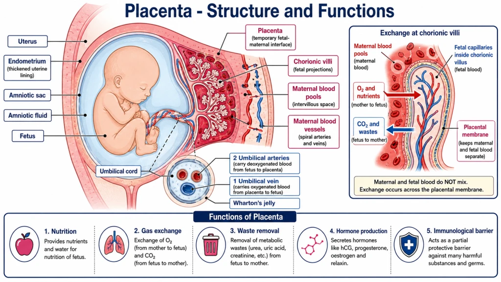
The placenta is a disc-shaped organ that:
- Connects the fetus to the uterine wall.
- Provides nutrition, gas exchange, waste removal, and hormone production.
- Develops from BOTH the trophoblast (fetal contribution) and endometrium (maternal contribution).
Two parts of the placenta
| Part | Origin | Composition |
|---|---|---|
| Fetal part (chorion) | Trophoblast | Chorionic villi |
| Maternal part (decidua) | Endometrium | Modified uterine tissue |
Definition (NCERT-canonical)
Placenta = a structural and functional unit between the developing embryo (fetus) and maternal body, formed from the chorionic villi (fetal) embedded in the maternal endometrium.
\
Functions of the Placenta
The placenta performs 5 critical functions for the developing fetus. Memorise all 5:
1. Nutrition
- Glucose, amino acids, fatty acids, vitamins, minerals pass from mother to fetus.
- Mostly via simple diffusion + active transport across placental membrane.
2. Gas exchange
- Oxygen (O₂) diffuses from maternal blood → fetal blood.
- Carbon dioxide (CO₂) diffuses from fetal blood → maternal blood.
- The placenta acts as the fetal lungs (since fetal lungs aren't functional in utero).
3. Waste removal
- Urea, uric acid, and other metabolic waste from the fetus are transferred to maternal blood.
- The mother's kidneys excrete these wastes through her urine.
- The placenta acts as the fetal kidneys.
4. Hormone production (endocrine function)
The placenta is itself an endocrine gland, producing several hormones:
- hCG (human Chorionic Gonadotropin) — first 3 months; maintains corpus luteum.
- hPL (human Placental Lactogen) — prepares mammary glands for lactation; alters maternal metabolism.
- Oestrogen (estriol mainly) — supports uterine growth, mammary preparation.
- Progesterone — maintains endometrium; suppresses uterine contractions.
- Relaxin (secreted by the ovary, not the placenta — NCERT) — relaxes the pelvic ligaments and softens the cervix (later in pregnancy).
After ~3 months, the placenta takes over hormone production from the corpus luteum.
5. Immunological barrier
- Prevents the mother's immune system from attacking the fetus (which is genetically half-foreign).
- Some maternal antibodies (IgG) can cross — providing passive immunity to the newborn.
What does NOT cross
- Most bacteria and large viruses are blocked.
- Maternal blood itself doesn't mix with fetal blood (they are separated by placental membrane).
- Some drugs and toxins can cross — important in pregnancy (alcohol, nicotine, certain medications must be avoided).
[NEET Important] The 5 functions: Nutrition + Gas exchange + Waste removal + Hormone production + Immunological barrier.
Pregnancy Hormones — A Cocktail of Change
Pregnancy is sustained by a cocktail of hormones, mostly produced by the placenta (after the corpus luteum's role declines).
1. hCG (human Chorionic Gonadotropin)
- Source: Trophoblast → later placenta.
- Time: Detectable from ~day 7-10; peaks at ~10 weeks; declines later.
- Function:
- Maintains corpus luteum for ~3 months (so progesterone continues).
- Basis of pregnancy tests (detected in urine).
- Importance: Without hCG, pregnancy can't continue past the first cycle.
2. hPL (human Placental Lactogen, also called hCS)
- Source: Placenta.
- Function:
- Prepares mammary glands for lactation.
- Alters maternal metabolism to make more glucose available for fetus.
- Increases insulin resistance in mother (so glucose stays in blood for fetus).
3. Oestrogen (mainly Estriol)
- Source: First placenta + maternal adrenal glands; later mainly placenta.
- Function:
- Promotes uterine growth (uterus expands ~500-1000× during pregnancy).
- Prepares mammary glands.
- Promotes maternal blood vessel development.
4. Progesterone
- Source: First corpus luteum (3 months); later placenta.
- Function:
- Maintains endometrium (key for pregnancy).
- Suppresses uterine contractions (prevents premature labour).
- Suppresses ovulation (no new cycles during pregnancy).
5. Relaxin (from the ovary, later in pregnancy)
- Source: the ovary, in the later phase of pregnancy (NCERT).
- Function:
- Relaxes pelvic ligaments to allow childbirth.
- Softens the cervix to prepare for dilation during labour.
Summary table
| Hormone | Source | Function |
|---|---|---|
| hCG | Trophoblast → placenta | Maintains corpus luteum (3 months); pregnancy test |
| hPL | Placenta | Mammary preparation; metabolic shifts |
| Oestrogen | Placenta | Uterine growth; mammary preparation |
| Progesterone | Corpus luteum → placenta | Endometrium maintenance; suppress contractions |
| Relaxin | Ovary (later phase of pregnancy) | Relaxes pelvic ligaments; softens cervix |
[NEET Important] hCG, hPL and relaxin are produced in women only during pregnancy; estrogens, progestogens, cortisol, prolactin and thyroxine rise several-fold in maternal blood (NCERT). hCG is the most-tested hormone of this group.
Small Memory Capsule
Section 12 in 60 seconds.
Placenta = mother-fetus interface
- Origin: Trophoblast (fetal) + endometrium (maternal)
- Two parts: Chorion (fetal) + Decidua (maternal)
- Connection: Fetus connects via umbilical cord (2 umbilical arteries + 1 umbilical vein)
5 functions
- Nutrition — glucose, amino acids, etc. cross to fetus
- Gas exchange — O₂ in, CO₂ out (placenta = fetal lungs)
- Waste removal — urea, uric acid (placenta = fetal kidneys)
- Hormone production — hCG, hPL, oestrogen and progesterone — while relaxin comes from the ovary in late pregnancy
- Immunological barrier — protects fetus; maternal antibodies cross (passive immunity)
5 pregnancy hormones (memorise!)
| Hormone | Source | Key function |
|---|---|---|
| hCG | Trophoblast/placenta | Maintains corpus luteum; pregnancy test |
| hPL | Placenta | Mammary preparation + metabolic shift |
| Oestrogen | Placenta | Uterine growth |
| Progesterone | Corpus luteum/placenta | Maintain endometrium; suppress contractions |
| Relaxin | Ovary (late pregnancy) | Relax pelvic ligaments; soften cervix |
Hormone timing
- First 3 months: corpus luteum is main hormone source (under hCG support).
- After 3 months: placenta takes over all hormone production.
- hCG peaks ~10 weeks, then declines.
- Estrogen + progesterone rise throughout pregnancy.
Maternal-fetal blood
- DO NOT mix directly!
- Separated by placental membrane.
- Exchange via diffusion + active transport.
One-line takeaway
"Placenta = temporary organ from chorion + decidua; provides nutrition, gas exchange, waste removal, hormone production (hCG, hPL, oestrogen and progesterone — while relaxin comes from the ovary in late pregnancy), and acts as immunological barrier; maternal and fetal blood don't mix but exchange substances across the placental membrane."
Solved Examples
Example 1: Define placenta
What is the placenta? Where does it come from?
Solution:
The placenta is a disc-shaped, temporary organ that forms during pregnancy as the interface between the developing fetus and the maternal body.
Origin:
- Fetal part (Chorion): From the trophoblast of the blastocyst. Forms chorionic villi.
- Maternal part (Decidua): From the endometrium of the uterus. Modified during pregnancy.
The placenta develops over the first 3 months of pregnancy and remains active until birth, when it's delivered as the afterbirth.
Answer: Placenta = mother-fetus interface; fetal chorion + maternal decidua.
Example 2: 5 functions
List the five major functions of the placenta.
Solution:
Nutrition — transfer of glucose, amino acids, fatty acids, vitamins, minerals from mother to fetus.
Gas exchange — O₂ from mother → fetus; CO₂ from fetus → mother (placenta = "fetal lungs").
Waste removal — urea, uric acid, etc. from fetus → mother for excretion (placenta = "fetal kidneys").
Hormone production — hCG, hPL, oestrogen and progesterone — while relaxin comes from the ovary in late pregnancy.
Immunological barrier — protects fetus from infection while allowing some maternal antibodies (IgG) to cross for passive immunity.
Answer: Nutrition + Gas exchange + Waste removal + Hormones + Immunological barrier.
Example 3: Hormone identification
What hormone is detected by urinary pregnancy tests? Where is it produced?
Solution:
hCG (human Chorionic Gonadotropin) is detected by urinary pregnancy tests.
- Production: Trophoblast cells of the blastocyst (and later the placenta).
- First detectable: ~7-10 days post-fertilisation in blood; ~10-14 days in urine.
- Function: Maintains the corpus luteum for ~3 months → progesterone continues → endometrium maintained → pregnancy continues.
- Peak: ~10 weeks of pregnancy.
- Decline: After 10 weeks, placenta takes over hormone production from corpus luteum, hCG levels decline.
A positive urine pregnancy test detects hCG → confirms the embryo has implanted.
Answer: hCG; produced by trophoblast/placenta; basis of pregnancy tests.
Example 4: Placental hormones
Name 4 hormones produced by the placenta and their functions.
Solution:
- hCG — Maintains corpus luteum (~3 months); basis of pregnancy tests.
- hPL (human Placental Lactogen) — Prepares mammary glands for lactation; alters maternal metabolism (increases glucose availability for fetus).
- Oestrogen — Promotes uterine growth (~500-1000× expansion); mammary preparation.
- Progesterone — Maintains endometrium; suppresses uterine contractions (preventing premature labour).
- Relaxin (from the ovary, late pregnancy — NCERT) — relaxes pelvic ligaments and softens the cervix.
Answer: hCG, hPL, oestrogen and progesterone — while relaxin comes from the ovary in late pregnancy (5 hormones; can also list 4).
Example 5: Maternal-fetal blood mixing
Do maternal and fetal blood mix in the placenta?
Solution:
NO — maternal and fetal blood DO NOT mix.
They are separated by the placental membrane (also called the placental barrier), which is composed of:
- Fetal capillary endothelium
- Connective tissue (fetal)
- Trophoblast (fetal)
- Maternal blood pools (in chorionic spaces)
Exchange occurs across this membrane by:
- Diffusion (gases, small molecules)
- Active transport (glucose, amino acids, ions)
- Endocytosis/exocytosis (some larger molecules)
Why is this important?
If maternal and fetal blood mixed:
- Blood type compatibility issues (e.g., Rh incompatibility could be catastrophic).
- Maternal immune cells would attack the fetus.
- Pathogens could pass directly.
The separation by the placental membrane allows essential exchange while maintaining biological compatibility.
Answer: No, they don't mix. Separated by placental membrane; only specific substances cross via diffusion/active transport.
Example 6: Why does pregnancy stop menstruation?
Why doesn't a pregnant woman menstruate?
Solution:
Pregnancy stops menstruation through a hormone cascade:
- Implanted blastocyst's trophoblast produces hCG.
- hCG maintains the corpus luteum — so it doesn't degenerate at day 28 as it normally would.
- Corpus luteum continues producing progesterone for ~3 months.
- Progesterone maintains the endometrium — so it doesn't shed.
- No menstruation occurs.
After ~3 months, the placenta takes over progesterone production. Endometrium continues to be maintained throughout pregnancy.
So the first sign of pregnancy for many women is a missed period (~day 32-35 of cycle), because the cycle that should have ended in menstruation continues into pregnancy.
Answer: hCG → maintains corpus luteum → progesterone continues → endometrium not shed → no menstruation throughout pregnancy.