
Grid 1 - The Brain's Control Centres, All in One Place
The chapter tells you what each part of the brain does, but it tells you in five separate places - once in the opening list of what the brain controls, once in the forebrain, once in the midbrain, once in the hindbrain, and once more in the summary. Nowhere does it put the whole thing in one table. That table is what this section is: an address grid, so that when an exam item names a function you can go straight to the part.
Nothing here is new. Every row is a fact already established elsewhere in this chapter.
| Part of the brain | What it controls |
|---|---|
| Cerebrum / cerebral cortex | Voluntary movements, and the processing of vision, hearing, speech, memory, intelligence, emotions and thoughts; its association areas handle intersensory associations, memory and communication |
| Thalamus | A major coordinating centre for sensory and motor signalling |
| Hypothalamus | Body temperature; the urge for eating and drinking; secretion of hypothalamic hormones from its neurosecretory cells; the circadian (24-hour) rhythms |
| Limbic system, along with the hypothalamus | Regulation of sexual behaviour, the expression of emotional reactions such as excitement, pleasure, rage and fear, motivation, and also olfaction and autonomic responses |
| Midbrain | Receives and integrates visual, tactile and auditory inputs |
| Cerebellum | The balance of the body, by integrating information from the semicircular canals of the ear and the auditory system |
| Pons | Its fibre tracts interconnect different regions of the brain |
| Medulla | Respiration, cardiovascular reflexes and gastric secretions |
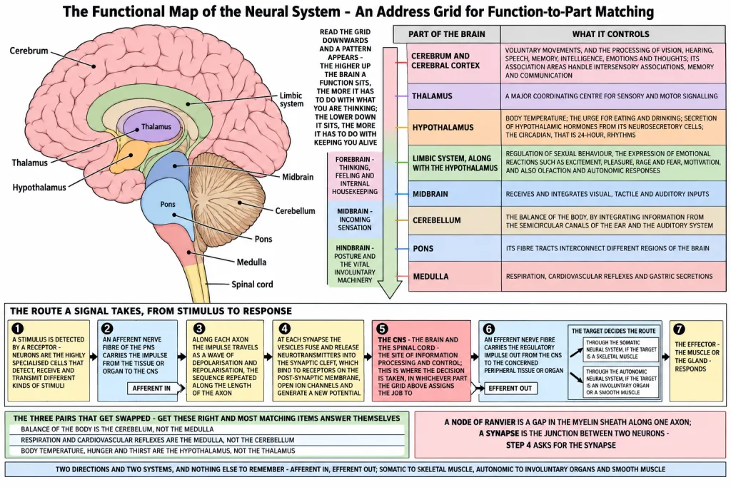
Read the grid downwards once and a pattern appears. The forebrain parts at the top handle the things you would call thinking, feeling and internal housekeeping. The midbrain in the middle handles incoming sensation. The hindbrain parts at the bottom handle posture and the vital involuntary machinery. The higher up the brain a function sits, the more it has to do with what you are thinking; the lower down it sits, the more it has to do with keeping you alive.
[NEET Important] Function-to-part matching is the commonest question type in this chapter, and the three pairs that get swapped are these: balance of the body is the cerebellum, not the medulla; respiration and cardiovascular reflexes are the medulla, not the cerebellum; and body temperature, hunger and thirst are the hypothalamus, not the thalamus. Get those three right and most of the matching items answer themselves.
Grid 2 - The Route a Signal Takes, from Stimulus to Response
The second thing the chapter never assembles is the journey. It describes the pieces - fibre types, impulse conduction, the synapse, the CNS - in separate sections, and never runs a single signal from one end to the other. Here is that run, built only from what the chapter has already stated:
- A stimulus is applied and is detected by a receptor. Neurons are the highly specialised cells that detect, receive and transmit different kinds of stimuli.
- An afferent nerve fibre of the peripheral neural system (PNS) carries the impulse from the tissue or organ TO the central neural system (CNS).
- Along each axon the impulse travels as a wave of depolarisation and repolarisation - the site ahead is depolarised in turn, and the sequence is repeated along the length of the axon so that the impulse is conducted.
- Wherever one neuron ends and the next begins, the impulse must cross a synapse. At a chemical synapse the arriving impulse moves the synaptic vesicles to the membrane, they fuse with it and release neurotransmitters into the synaptic cleft; these bind to specific receptors on the post-synaptic membrane, which opens ion channels and generates a new potential in the next neuron.
- The impulse reaches the CNS - the brain and the spinal cord - which is the site of information processing and control. This is where the decision is taken, in whichever part of the brain Grid 1 assigns the job to.
- An efferent nerve fibre carries the regulatory impulse out from the CNS to the concerned peripheral tissue or organ. Which route it takes depends on the target:
- through the somatic neural system, if the target is a skeletal muscle;
- through the autonomic neural system, if the target is an involuntary organ or a smooth muscle.
- The effector - the muscle or the gland - responds, and that response is the body's answer to the stimulus in step 1.
Two directions and two systems, and nothing else to remember: afferent in, efferent out; somatic to skeletal muscle, autonomic to involuntary organs and smooth muscle.
[NEET Important] Items on this route usually attack one join. The join between step 2 and step 6 is afferent versus efferent - direction, and the word regulatory on the outgoing impulse. The join inside step 6 is somatic versus autonomic - the target decides. And step 4 is the one that asks for the synapse, not the node of Ranvier; a node is a gap in the myelin sheath along one axon, a synapse is the junction between two neurons.
Grid 3 - The Three Divisions Against the Three Regions of the Brain Stem
The last thing to line up is the pair of overlapping schemes. The brain is divided into three major parts - forebrain, midbrain and hindbrain. Separately, three major regions make up the brain stem - midbrain, pons and medulla oblongata. Those are two different ways of cutting up the same brain, and they do not agree.
| Division of the brain | Parts of that division | Part of the brain stem? |
|---|---|---|
| Forebrain | Cerebrum | No |
| Forebrain | Thalamus | No |
| Forebrain | Hypothalamus | No |
| Midbrain | Midbrain | Yes - wholly |
| Hindbrain | Pons | Yes |
| Hindbrain | Medulla oblongata | Yes |
| Hindbrain | Cerebellum | No |
Three readings of that grid:
- The forebrain contributes nothing at all to the brain stem. Cerebrum, thalamus and hypothalamus are all outside it.
- The midbrain is wholly inside the brain stem - it is the only division that is entirely part of it.
- The hindbrain is split: the pons and the medulla oblongata are in the brain stem, the cerebellum is not.
And the reason for the odd grouping is functional, not anatomical tidiness: the brain stem forms the connections between the brain and the spinal cord, so it collects the parts that lie on that connecting stalk, wherever they happen to fall in the three-division scheme.
[NEET Important] The single most-missed fact in this chapter: the cerebellum is part of the hindbrain but NOT part of the brain stem. The second: the brain stem is not a fourth division of the brain - the divisions are forebrain, midbrain and hindbrain, and the brain stem is a grouping laid across two of them.
Quick Recap
- The cerebrum controls voluntary movements and is the site for processing vision, hearing, speech, memory, intelligence, emotions and thoughts; its association areas handle intersensory associations, memory and communication.
- The thalamus is the major coordinating centre for sensory and motor signalling.
- The hypothalamus controls body temperature and the urge for eating and drinking, secretes hypothalamic hormones, and runs the circadian (24-hour) rhythms.
- The limbic system along with the hypothalamus handles sexual behaviour, the expression of emotional reactions, motivation, olfaction and autonomic responses.
- The midbrain receives and integrates visual, tactile and auditory inputs.
- The cerebellum looks after the balance of the body and integrates information from the semicircular canals of the ear and the auditory system.
- The pons consists of fibre tracts that interconnect different regions of the brain.
- The medulla contains centres controlling respiration, cardiovascular reflexes and gastric secretions.
- The route of a signal: receptor - afferent fibre - CNS (site of information processing and control)
- efferent fibre - somatic to skeletal muscle or autonomic to involuntary organ and smooth muscle - effector.
- Along an axon the impulse is a wave of depolarisation and repolarisation; between two neurons it crosses a synapse.
- The three major parts of the brain are forebrain, midbrain and hindbrain; the three regions of the brain stem are midbrain, pons and medulla oblongata.
- The forebrain contributes nothing to the brain stem, the midbrain is wholly in it, and of the hindbrain the pons and medulla are in it while the cerebellum is not.
Solved Examples
Question 1
Q. Differentiate between the cerebrum and the cerebellum. This is one of the chapter-end exercises.
Answer.
| Feature | Cerebrum | Cerebellum |
|---|---|---|
| Division of the brain | Forebrain | Hindbrain |
| Relative size | Forms the major part of the human brain - the largest part | Much smaller, lying below and behind the cerebrum |
| Surface | Thrown into prominent folds, and divided by a deep cleft into left and right cerebral hemispheres joined by the corpus callosum | Very convoluted, in order to provide the additional space for many more neurons |
| Name of the surface layer | The cerebral cortex, called the grey matter because of the concentration of neuron cell bodies | No such named cortex is given in this chapter |
| Function | Voluntary movements, and the processing of vision, hearing, speech, memory, intelligence, emotions and thoughts; its association areas handle intersensory associations, memory and communication | The balance of the body; it integrates information received from the semicircular canals of the ear and the auditory system |
| Part of the brain stem? | No | No |
The one line that gets the mark: the cerebrum is the largest part of the brain, belonging to the forebrain, and is the seat of voluntary action, thought and the special senses, while the cerebellum is a hindbrain part that maintains the balance of the body.
Question 2
Q. Make a table naming the part of the brain that controls each of the following: body temperature, balance of the body, respiration, sensory and motor coordination, and visual and auditory input.
Answer.
| Function | Part of the brain |
|---|---|
| Body temperature | Hypothalamus |
| Balance of the body | Cerebellum |
| Respiration | Medulla |
| Sensory and motor coordination | Thalamus |
| Visual, tactile and auditory input | Midbrain |
Question 3
Q. Trace the route taken by a signal from the moment a stimulus is applied to the moment the body responds, naming the fibres and the systems involved.
Answer. In order:
- The stimulus is detected by a receptor - neurons detect, receive and transmit different kinds of stimuli.
- An afferent nerve fibre of the PNS carries the impulse from the tissue or organ to the CNS.
- Along the axon the impulse travels as a wave of depolarisation and repolarisation, the sequence being repeated along the length of the fibre.
- At each junction between two neurons the impulse crosses a synapse; at a chemical synapse neurotransmitters are released into the synaptic cleft, they bind to receptors on the post-synaptic membrane, and a new potential is generated in the next neuron.
- The impulse reaches the CNS - the brain and the spinal cord - the site of information processing and control, where it is processed.
- An efferent nerve fibre carries the regulatory impulse from the CNS to the concerned peripheral tissue or organ: through the somatic neural system to a skeletal muscle, or through the autonomic neural system to an involuntary organ or smooth muscle.
- The effector responds.
Question 4
Q. Which division of the brain contributes nothing to the brain stem, and which division lies wholly within it?
Answer. The forebrain contributes nothing to the brain stem - the cerebrum, thalamus and hypothalamus are all outside it. The midbrain lies wholly within the brain stem. Of the hindbrain, the pons and the medulla oblongata are in the brain stem while the cerebellum is not.
Question 5
Q. Which part of the brain would you hold responsible for a rise in body temperature going uncorrected, and why?
Answer. The hypothalamus, because it contains a number of centres which control body temperature as well as the urge for eating and drinking.
Question 6
Q. A person can walk, but staggers and cannot keep their balance. Which part of the brain is likely affected, and what does that part normally do?
Answer. The cerebellum. It looks after the balance of the body and integrates information received from the semicircular canals of the ear and the auditory system - the very information needed to stay upright.
Question 7
Q. Name the part of the brain that houses the centres for respiration, cardiovascular reflexes and gastric secretions.
Answer. The medulla, also called the medulla oblongata, which is connected to the spinal cord.
Question 8
Q. Which part of the brain is the master clock, and which part is the major coordinating centre for sensory and motor signalling?
Answer. The hypothalamus is the master clock, because it runs the body's circadian (24-hour) rhythms. The thalamus is the major coordinating centre for sensory and motor signalling.
Question 9
Q. Which part of the brain receives and integrates visual, tactile and auditory inputs, and where does it lie?
Answer. The midbrain. It lies between the thalamus/hypothalamus of the forebrain and the pons of the hindbrain.
Question 10
Q. Which structures of the brain are involved in olfaction and in the expression of emotional reactions?
Answer. The limbic system, along with the hypothalamus. Between them they handle olfaction, autonomic responses, the regulation of sexual behaviour, the expression of emotional reactions such as excitement, pleasure, rage and fear, and motivation.
Question 11
Q. Which part of the brain consists of fibre tracts that interconnect different regions of the brain, and which division does it belong to?
Answer. The pons, which belongs to the hindbrain. It is also one of the three regions of the brain stem.
Question 12
Q. An impulse must reach a skeletal muscle of the arm and, at the same time, the smooth muscle of the gut. Name the division of the PNS used in each case.
Answer. For the skeletal muscle of the arm, the somatic neural system, which relays impulses from the CNS to the skeletal muscles. For the smooth muscle of the gut, the autonomic neural system, which transmits impulses from the CNS to the involuntary organs and smooth muscles of the body.
Question 13
Q. Fill in the blanks: the impulse travels along an axon as a wave of _ and , crosses the junction between two neurons at a , is processed in the , and leaves along an _ fibre.
Answer. The impulse travels along an axon as a wave of depolarisation and repolarisation, crosses the junction between two neurons at a synapse, is processed in the CNS, and leaves along an efferent fibre.